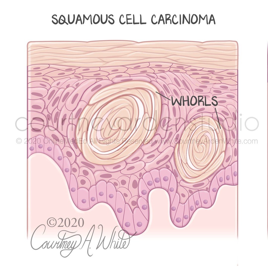
This illustration was created for medical education about the histological characteristics of skin cancer as seen under the microscope. The three major types are squamous cell carcinoma, basal cell carcinoma, and Melanoma. Squamous cell carcinoma is composed of spindle cells in a whorled pattern. Basal cell carcinoma is characterized by basaloid nests that stain more blue. Melanoma appears as nested melanocytes of varying sizes.
#medicalillustration #illustrationsmedical #medart #medicalartwork #sciart #biologicalart #medicalcommunication #anatomyart #anatomicalart #artandanatomy #anatomydrawing #biologicalillustration #anatomyillustration #drawinganatomy #scienceart #scienceartist #scienceandart #medicalartist #medicaleducation #medicalstudent #meded #scicomm #scicommunity #healthcareart #patienteducation #skincancer #skinhealth #melanomaawareness #skincheck #courtneyardenstudio
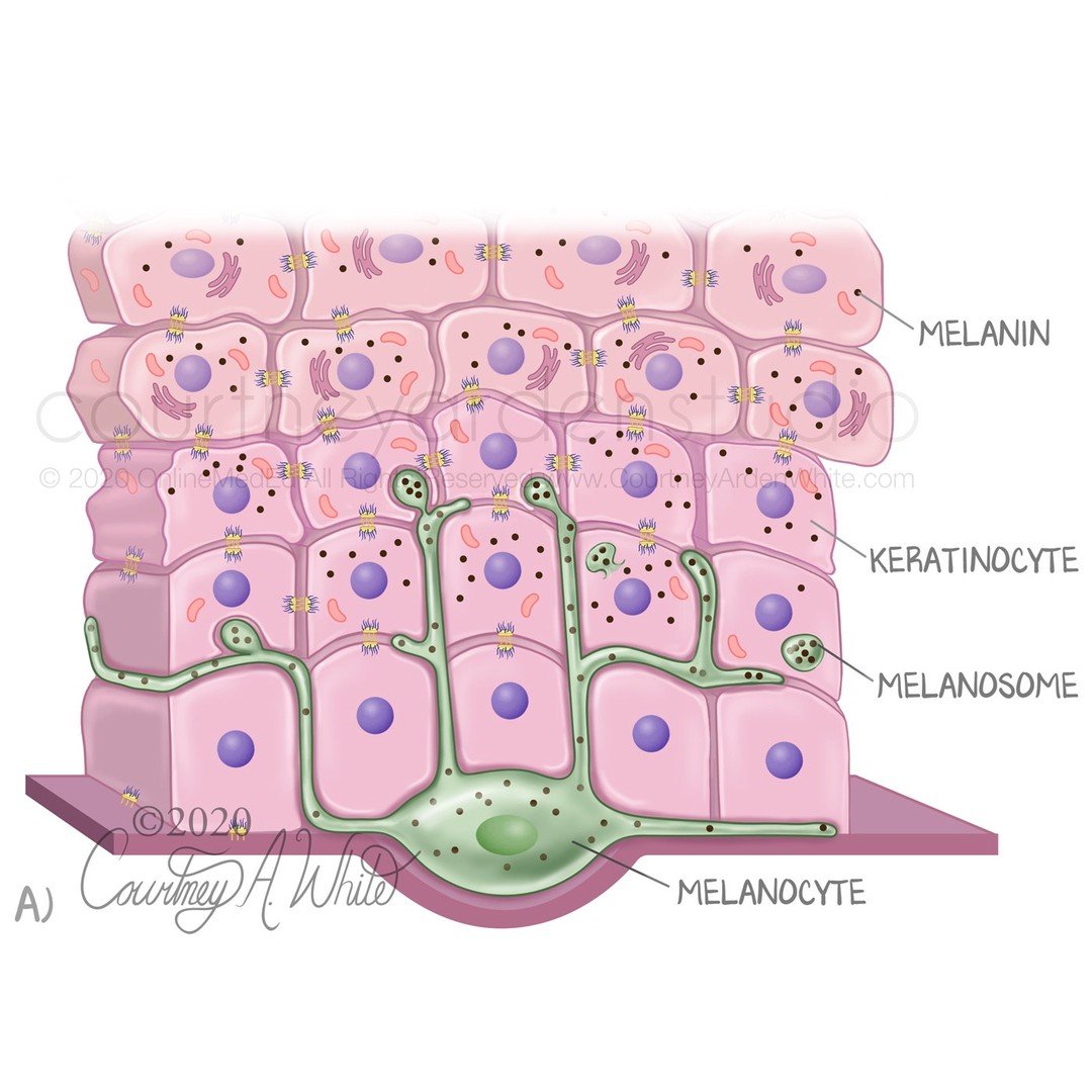
This illustration was created for medical education to explain the role of melanocytes in our body. Melanocytes are cells that produce the pigment melanin. Melanosomes transfer melanin to the surrounding keratinocytes. The melanin then organizes above the nucleus inside each cell and protects the skin from UV radiation. Additionally, melanocytes create pigmentation in the skin and hair. Although everyone has a similar number of melanocytes, the amount of melanin produced varies, resulting in different skin colors. When exposed to sunlight, melanin production increases, giving the appearance of a tan. Keratinocytes degrade the melanin more efficiently in light skin than in dark skin, which is what is shown on the right side of the illustration.
#medicalillustration #illustrationsmedical #medart #medicalartwork #sciart #biologicalart #medicalcommunication #anatomyart #anatomicalart #artandanatomy #anatomydrawing #biologicalillustration #anatomyillustration #drawinganatomy #scienceart #scienceartist #scienceandart #medicalartist #medicaleducation #medicalstudent #meded #scicommunity #healthcareart #patienteducation #melanocytes #skinillustrations #skinhealth #cellart #cellbiology #courtneyardenstudio
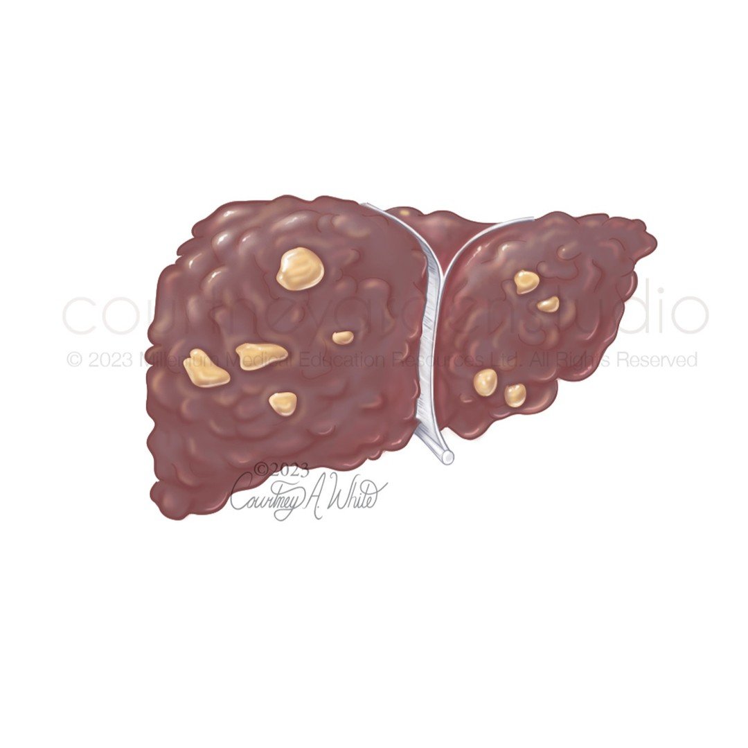
These illustrations were made for a patient education tear pad and tent card about the liver and liver cancer. I designed and wrote the content for this piece as well. Hepatocellular carcinoma (HCC) is the most prevalent type of liver cancer in adults, and it typically occurs in people with cirrhosis of the liver. HCC usually starts as several small cancerous lesions that spread throughout the liver.
#medicalillustration #illustrationsmedical #medart #medicalartwork #sciart #biologicalart #medicalcommunication #anatomyart #anatomicalart #artandanatomy #anatomydrawing #biologicalillustration #anatomyillustration #drawinganatomy #scienceart #scienceartist #scienceandart #medicalartist #medicaleducation #medicalstudent #meded #scicomm #scicommunity #healthcareart #patienteducation #Liverhealth #hepatocellularcarcinoma #liverdiseaseawareness #liverdisease #courtneyardenstudio
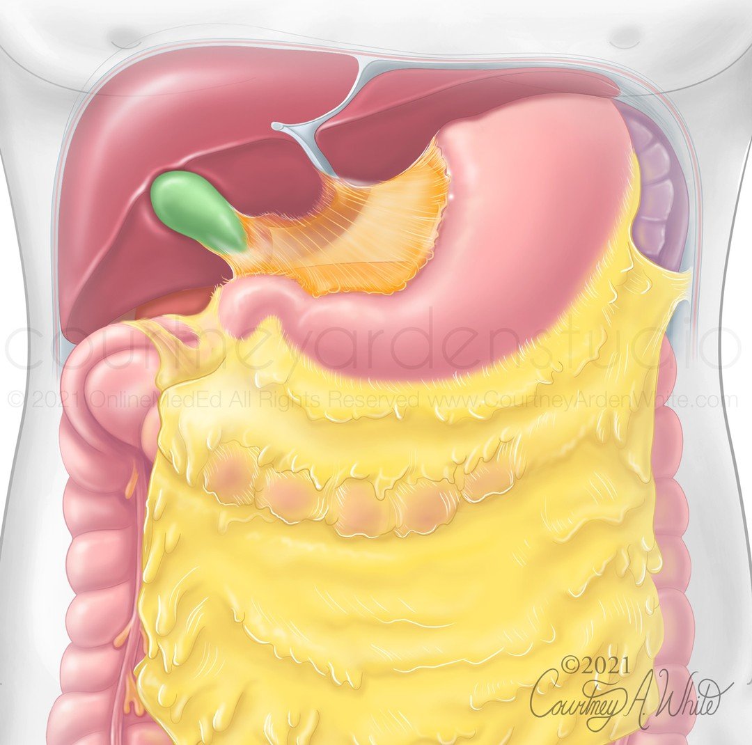
This illustration was created for medical education for medical students about the relative anatomy of the stomach. The stomach is located in the upper left quadrant of the abdomen. It is partially covered anteriorly by the liver and is located anterior to the pancreas and left kidney. The lesser curvature of the stomach is connected to the liver by the lesser omentum, and the greater curvature of the stomach is connected to the greater omentum, which drapes over the intestines.
#medicalillustration #illustrationsmedical #medart #medicalartwork #sciart #biologicalart #medicalcommunication #anatomyart #anatomicalart #artandanatomy #anatomydrawing #biologicalillustration #anatomyillustration #drawinganatomy #scienceart #scienceartist #scienceandart #medicalartist #medicaleducation #medicalstudent #meded #scicomm #scicommunity #healthcareart #patienteducation #gitracthealth #guthealth #gutanatomy #internalorgans #courtneyardenstudio
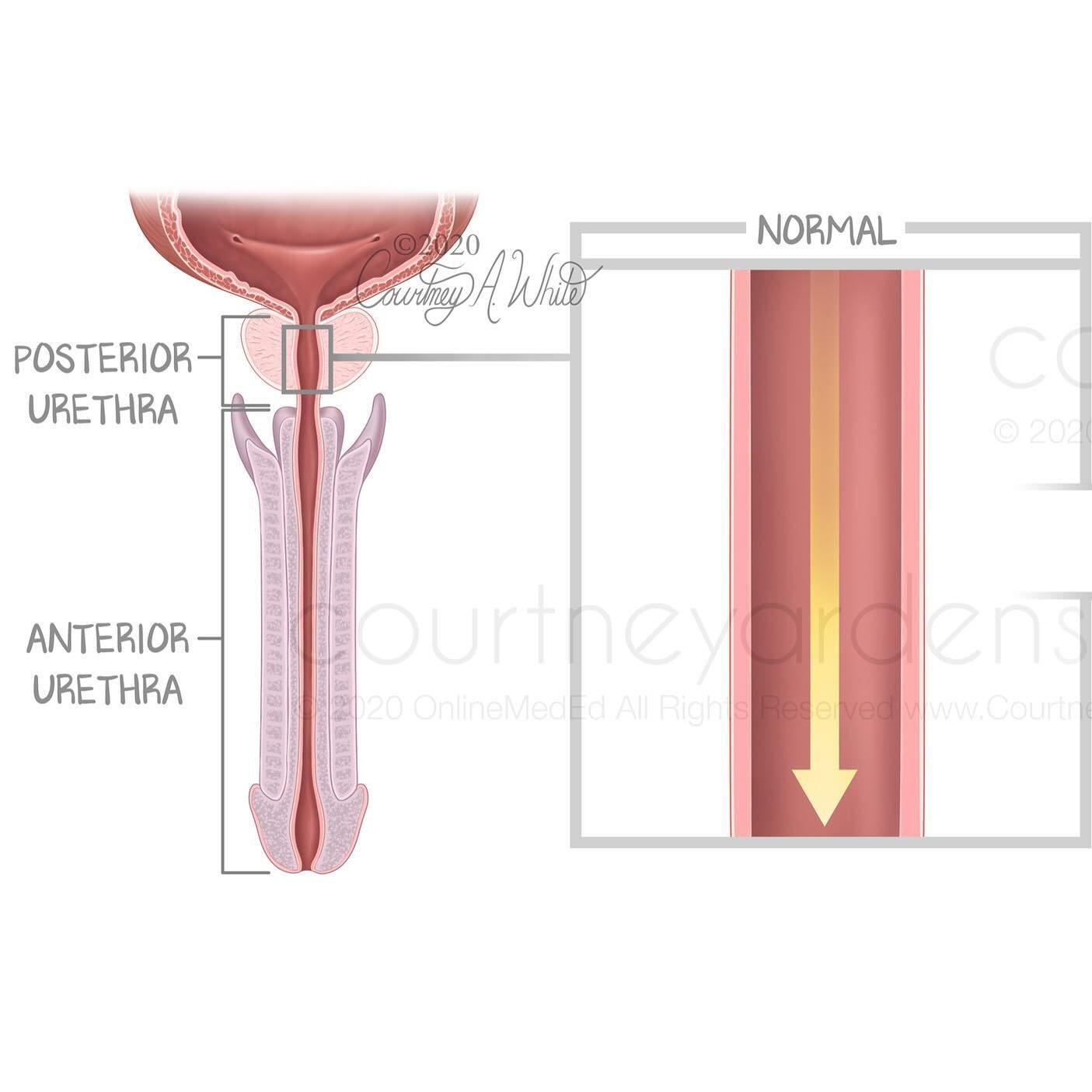
A posterior urethral valve (PUV) is a rare birth defect where a fold of tissue in the posterior part of the urethra obstructs the flow of urine. If urine isn’t able to leave the body it can damage the urethra, bladder, ureters and kidneys. The most common treatment is to surgically remove the tissue to clear the obstruction.
#medicalillustration #illustrationsmedical #medart #medicalartwork #sciart #biologicalart #medicalcommunication #anatomyart #anatomicalart #artandanatomy #anatomydrawing #biologicalillustration #anatomyillustration #drawinganatomy #scienceart #scienceartist #scienceandart #medicalartist #medicaleducation #medicalstudent #meded #scicomm #scicommunity #healthcareart #patienteducation #posteriorurethralvalves #congenitaldefect #humandevelopment #birthdefects #courtneyardenstudio
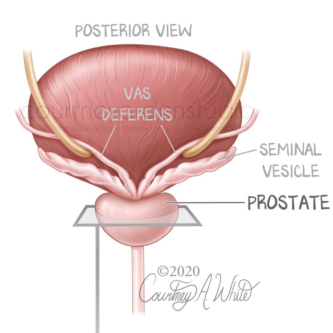
This illustration was made for medical education purposes regarding the prostate gland. The prostate gland is located below the bladder and in front of the rectum. It encircles the urethra, which is why an enlarged prostate due to conditions such as benign prostate hyperplasia or cancer can lead to disruptions in normal bladder habits.
#medicalillustration #illustrationsmedical #medart #medicalartwork #sciart #biologicalart #medicalcommunication #anatomyart #anatomicalart #artandanatomy #anatomydrawing #biologicalillustration #anatomyillustration #drawinganatomy #scienceart #scienceartist #scienceandart #medicalartist #medicaleducation #medicalstudent #meded #scicomm #scicommunity #healthcareart #patienteducation #prostategland #prostateenlargement #prostatehealth #prostate #courtneyardenstudio
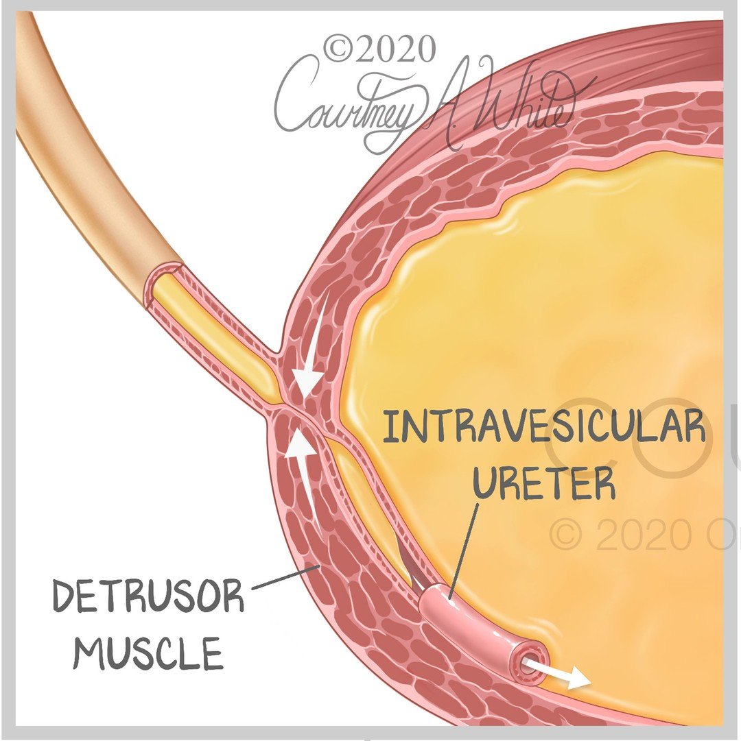
Vesicoureteral Reflex (VUR) is when urine flows backward from the bladder to the ureters and potentially to the kidneys. Normally, urine should flow from the kidneys, through the ureters, to the bladder. VUR can happen due to an abnormal vesicoureteral junction where the ureter connects to the bladder. Normally, there is an intravesicular portion of the ureter; however, if this is missing or is too short, it can cause this condition, which is mostly seen in infants and young children.
#medicalillustration #illustrationsmedical #medart #medicalartwork #sciart #biologicalart #medicalcommunication #anatomyart #anatomicalart #artandanatomy #anatomydrawing #biologicalillustration #anatomyillustration #drawinganatomy #scienceart #scienceartist #scienceandart #medicalartist #medicaleducation #medicalstudent #meded #scicomm #scicommunity #healthcareart #urologyhealthcare #bladderhealth #bladderproblems #kidneyhealth #patienteducation #courtneyardenstudio
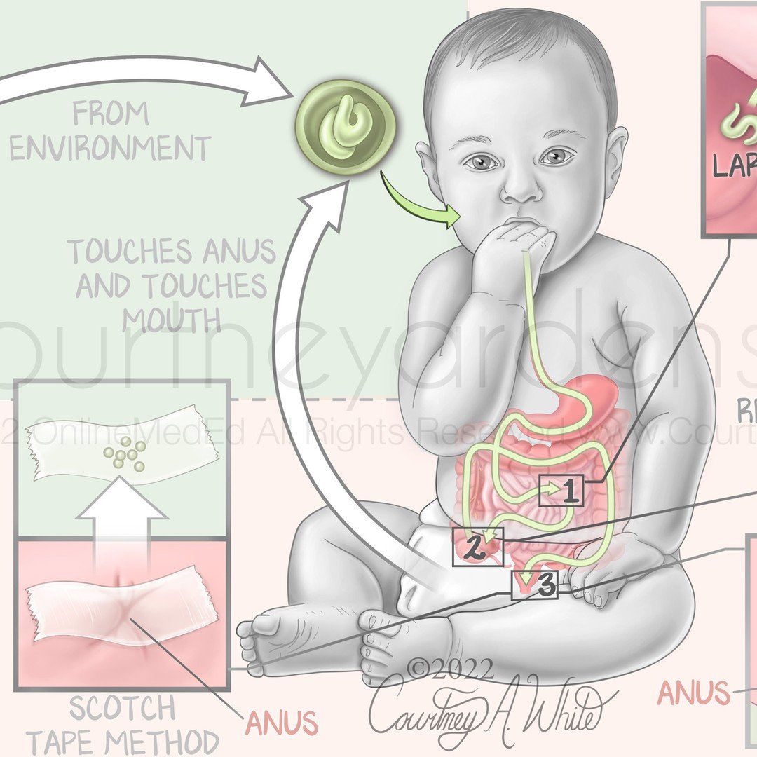
This medical illustration is part of a series of illustrations designed for medical education on parasites. Enterobius vermicularis, also known as threadworm or pinworm, is a parasitic roundworm that mostly infects children and is one of the most common helminth infections worldwide. At night, adult worms lay eggs around the anus, which can be tested for by applying clear tape to the anus in the morning. The eggs will stick to the tape, and they can be viewed under a microscope. If the eggs are swallowed, re-infection can occur.
#medicalillustration #medart #medicalart #sciart #biologicalart #medicalcommunication #anatomyart #anatomicalart #artandanatomy #anatomydrawing #biologicalillustration #anatomyillustration #drawinganatomy #scienceart #scienceartist #scienceandart #medicalartist #medicaleducation #medicalstudent #meded #scicomm #healthcareart #patienteducation #pinworms #enterobiusvermicularis #parasites #parasiticworms #helminth #illustrationsmedical #courtneyardenstudio
These illustrations were created for a two-page patient education tear pad on metastatic triple-negative breast cancer (mTNBC). The term triple-negative refers to three receptors commonly found in breast cancer: the estrogen receptor (ER), progesterone receptor (PR), and human epidermal growth factor receptor 2 (HER2). If the cancer cells don't express these, it is called triple negative.
#medicalillustration #medart #medicalart #sciart #biologicalart #medicalcommunication #anatomyart #anatomicalart #artandanatomy #anatomydrawing #biologicalillustration #anatomyillustration #drawinganatomy #scienceart #scienceartist #scienceandart #medicalartist #medicaleducation #medicalstudent #meded #scicomm #scicommunity #healthcareart #patienteducation #mtnbc #breastcancer #cellularart #cells #metastatictnbc #courtneyardenstudio
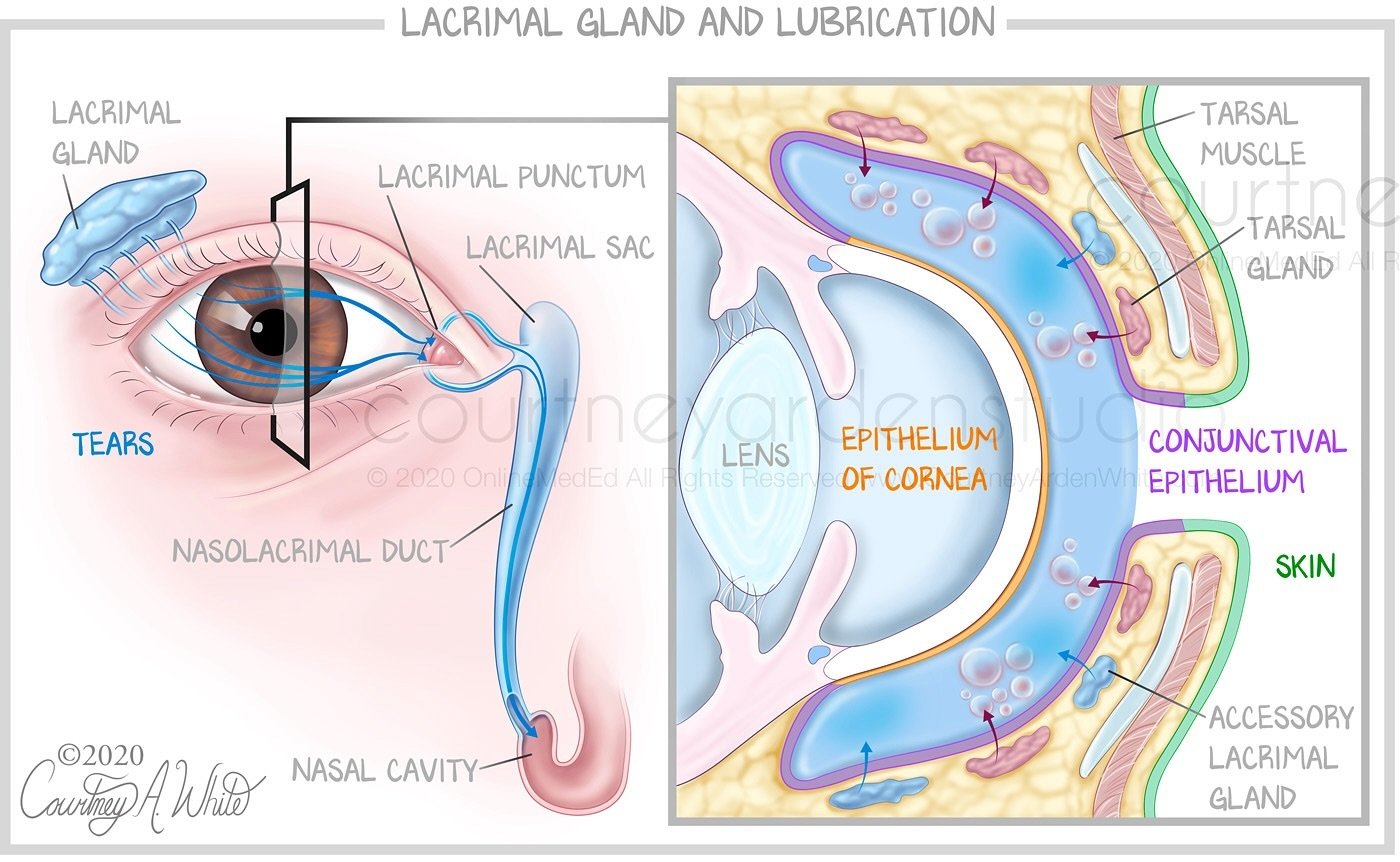
The lacrimal gland secretes tears to lubricate and protect the eyes. The excess tears drain into the nasolacrimal duct that connects to the nasal cavity. This medical illustration was created for medical education.
#medicalillustration #medart #medicalart #sciart #biologicalart #medicalcommunication #anatomyart #anatomicalart #artandanatomy #anatomydrawing #biologicalillustration #anatomyillustration #drawinganatomy #scienceart #scienceartist #scienceandart #medicalartist #medicaleducation #medicalstudent #meded #scicomm #scicommunity #healthcareart #patienteducation #eyehealth #lacrimalsurgery #eyeanatomyandphysiology #eyeanatomy #eyesurgery #courtneyardenstudio
In celebration of the solar eclipse, I wanted to highlight the eye, which is full of intricate muscles and cells that we don't want to damage. The rectus and oblique muscles of the eye allow the eye to rotate in each direction. The cells of the eye are critical for our ability to see. When light enters the eye, passing through the lens, it focuses on the retina in the back of the eye, where the photoreceptor cells consisting of rods and cones absorb the light. Then, the bipolar cells send the visual signal to the ganglion cells, and they convey the information to the brain. You can damage these important cells if you stare at the eclipse without the proper eyewear.
#medicalillustration #medart #medicalart #sciart #biologicalart #medicalcommunication #anatomyart #anatomicalart #artandanatomy #anatomydrawing #biologicalillustration #anatomyillustration #drawinganatomy #scienceart #scienceartist #scienceandart #medicalartist #medicaleducation #medicalstudent #meded #healthcareart #patienteducation #solareclipseeyes #solareclipse2024 #eyecells #retinadamage #solareclipseeyewear #eyehealthtips #eyesafety #courtneyardenstudio
This is another illustration about the GI tract that was created for medical education. It shows where different nutrients are absorbed in the digestive tract. Most absorption occurs in the small intestine, which consists of three parts: the duodenum, the jejunum, and the ileum. After the ileum comes the large intestine, where any remaining bile acids that were not absorbed in the ileum are absorbed.
#medicalillustration #medart #medicalart #sciart #biologicalart #medicalcommunication #anatomyart #anatomicalart #artandanatomy #anatomydrawing #biologicalillustration #anatomyillustration #drawinganatomy #scienceart #scienceartist #scienceandart #medicalartist #medicaleducation #medicalstudent #meded #scicomm #scicommunity #healthcareart #patienteducation #intestinalhealth #digestionhealth #digestivetract #gastrointestinal #digestionhealing #courtneyardenstudio
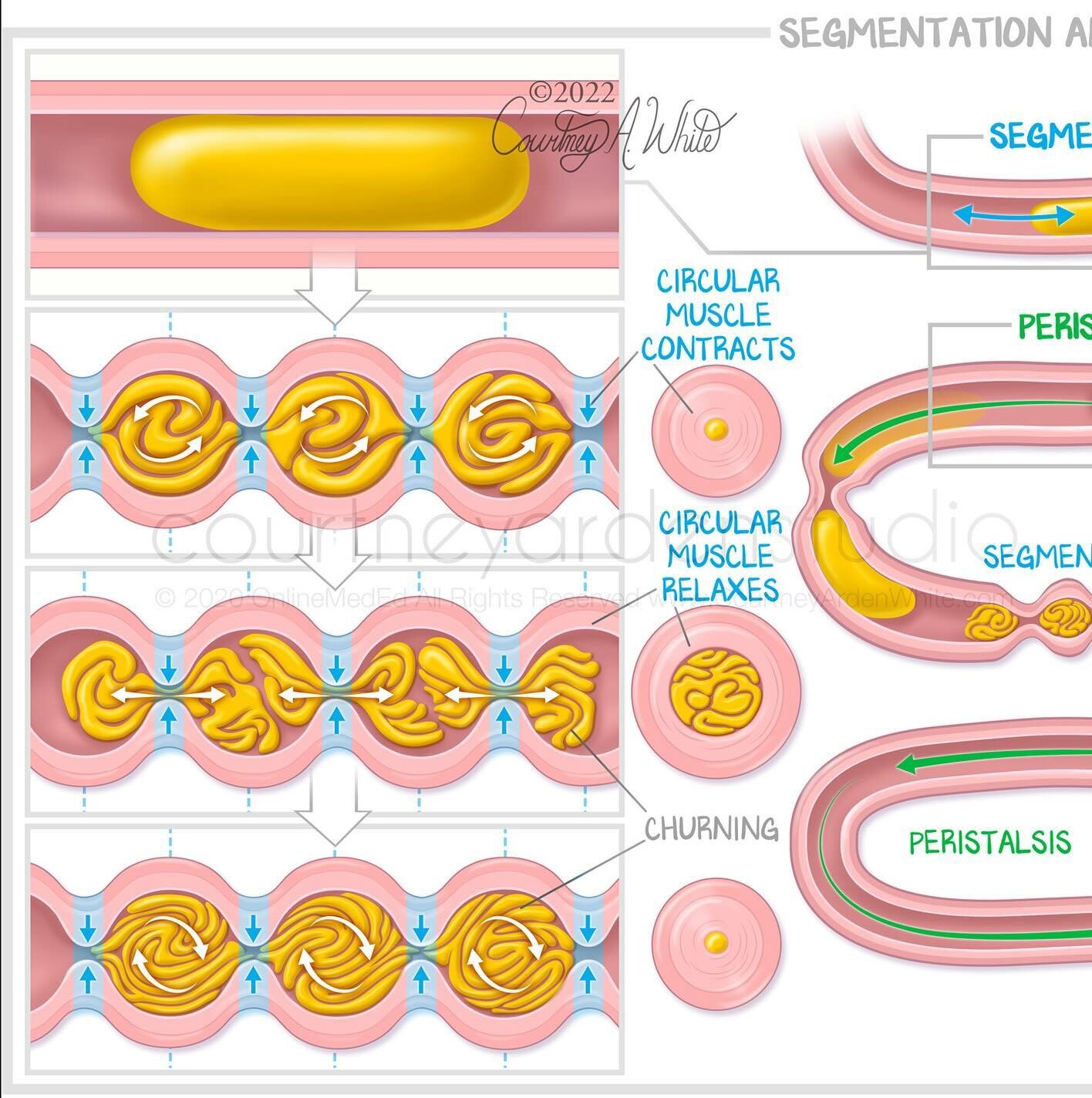
This illustration about segmentation and peristalsis of the small intestine was created for medical education. Segmentation and peristalsis are two ways the muscles of the small intestine moves the contents or chyme through the tract. Segmentation causes a mixing motion to allow for absorption of water and nutrients. Peristalsis causes the chyme to propel forward along the tract.
#medicalillustration #medart #medicalart #sciart #biologicalart #medicalcommunication #anatomyart #anatomicalart #artandanatomy #anatomydrawing #biologicalillustration #anatomyillustration #drawinganatomy #scienceart #scienceartist #scienceandart #medicalartist #medicaleducation #medicalstudent #meded #scicomm #scicommunity #healthcareart #patienteducation #peristalsis #digestionhealth #smallintestine #digestionanatomy #gitracthealth #courtneyardenstudio
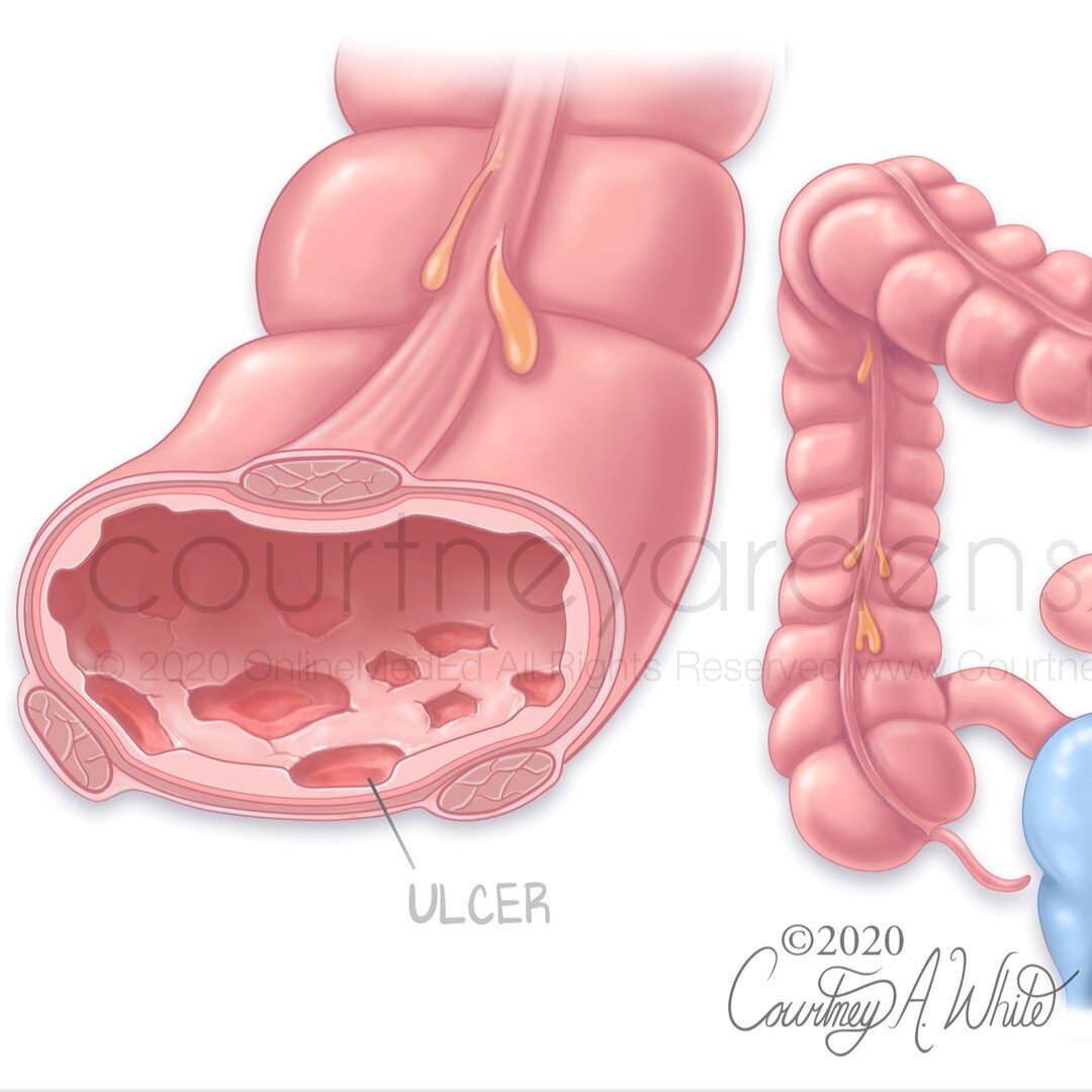
This medical illustration was designed for medical educational purposes to highlight the differences between ulcerative colitis and Crohn's disease, both of which are inflammatory bowel diseases (IBD). While Crohn's can cause inflammation anywhere in the gastrointestinal tract, including the mouth, ulcerative colitis only causes inflammation and ulceration in the large intestine. Additionally, ulcerative colitis affects only the inner layer of the large intestine wall, whereas Crohn's can reach all layers, as shown in the illustration by the knife-like fissures. Furthermore, ulcerative colitis causes continuous lesions or inflammation, while Crohn's has skip lesions where there will be a healthy part of the intestine between two inflamed parts.
#medicalillustration #medart #medicalart #sciart #biologicalart #medicalcommunication #anatomyart #anatomicalart #artandanatomy #anatomydrawing #biologicalillustration #anatomyillustration #drawinganatomy #scienceart #scienceartist #scienceandart #medicalartist #medicaleducation #medicalstudent #meded #scicomm #scicommunity #healthcareart #patienteducation #ulcerativecolitis #crohnsdisease #intestinalhealth #inflammatoryboweldisease #IBD #courtneyardenstudio
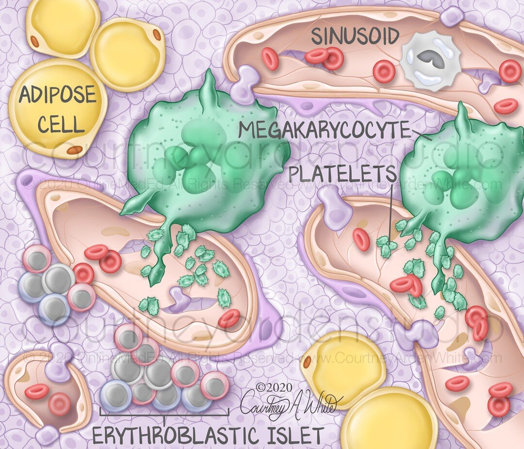
This illustration was created for medical education. It shows the histologic view of bone marrow and the relevant vocabulary. Blood cells are mostly made in the bone marrow and enter the bloodstream through sinusoids.
#medicalillustration #medart #medicalart #sciart #biologicalart #medicalcommunication #anatomyart #anatomicalart #artandanatomy #anatomydrawing #biologicalillustration #anatomyillustration #drawinganatomy #scienceart #scienceartist #scienceandart #medicalartist #medicaleducation #medicalstudent #meded #scicomm #scicommunity #healthcareart #patienteducation #bonemarrow #bonemarrowdonor #cellart #hematopoiesis #microscopic #courtneyardenstudio
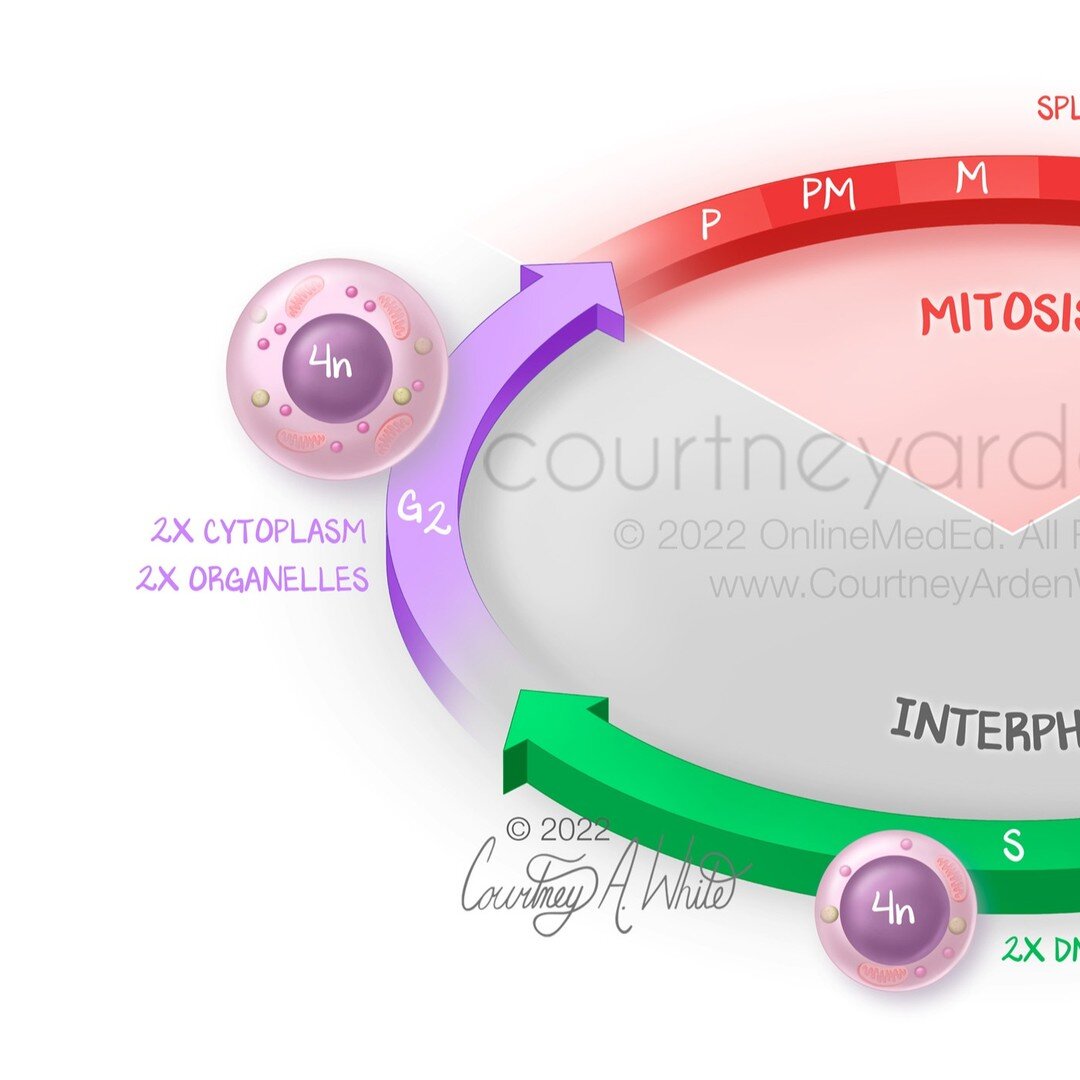
This is my last post about mitosis for the moment. This illustration shows the entire cell cycle, which can be divided into two main parts: interphase and mitosis. Most of the time, a cell is in interphase, where DNA replication and cell growth occur. Interphase can be divided further into G1 phase, S phase, and G2 phase. G1 and G2 are considered the gap phases. If the conditions around the cell aren't favorable for division, it may enter the G0 phase, where it could remain indefinitely or until the environment is more favorable. The S phase is when DNA replication occurs. After G2, the cell leaves interphase and enters mitosis, where the daughter chromosomes separate and usually end with the cells dividing.
#medicalillustration #medart #medicalart #sciart #biologicalart #medicalcommunication #anatomyart #anatomicalart #artandanatomy #anatomydrawing #biologicalillustration #anatomyillustration #drawinganatomy #scienceart #scienceartist #scienceandart #medicalartist #medicaleducation #medicalstudent #meded #scicomm #scicommunity #healthcareart #patienteducation #cellularart #mitosis #cellcycle #cellreplication #celldivision #courtneyardenstudio
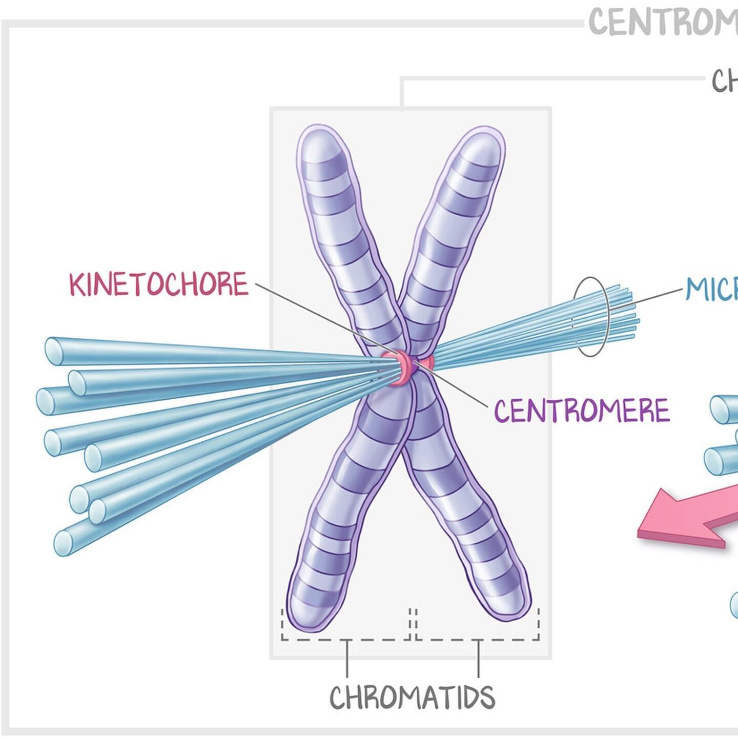
To continue with the mitosis illustrations, this one explains centromere vocabulary for medical education. The centromere is located between the two sister chromatids, holding them together. The kinetochore attaches the centromeres to the microtubules before cell division. Together, they power the movement of the chromosomes and separation into sister chromatids.
#medicalillustration #medart #medicalart #sciart #biologicalart #medicalcommunication #anatomyart #anatomicalart #artandanatomy #anatomydrawing #biologicalillustration #anatomyillustration #drawinganatomy #scienceart #scienceartist #scienceandart #medicalartist #medicaleducation #medicalstudent #meded #scicomm #scicommunity #healthcareart #cellularart #mitosis #kinetochore #centromere #microtubules #celldivision #courtneyardenstudio
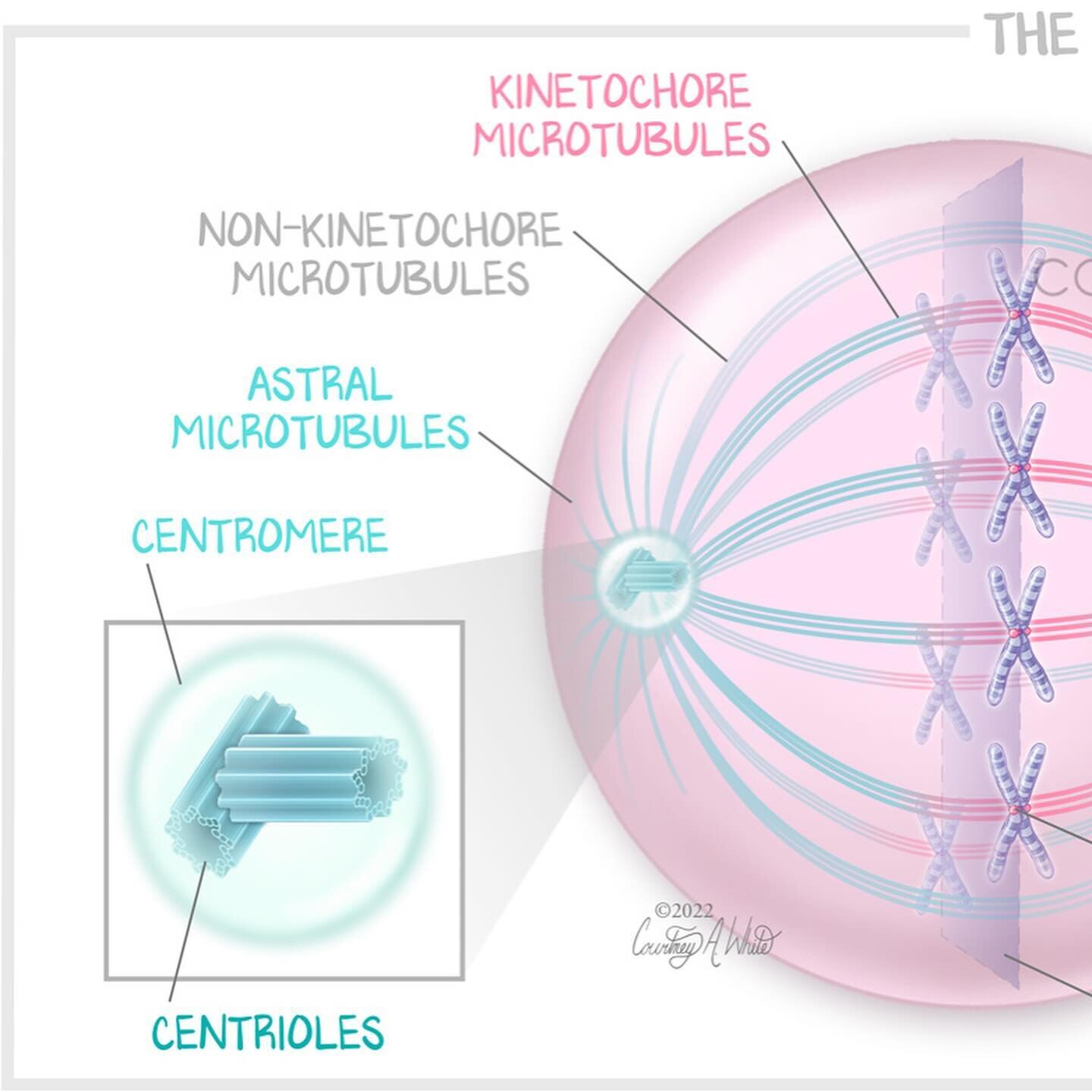
This illustration about the mitotic spindle was created for medical education. The mitotic spindle is an essential component of mitosis. It’s composed mostly of microtubules, and its job is to equally divide the chromosomes in the parent cell during cell division.
#medicalillustration #medart #medicalart #sciart #biologicalart #medicalcommunication #anatomyart #anatomicalart #artandanatomy #anatomydrawing #biologicalillustration #anatomyillustration #drawinganatomy #scienceart #scienceartist #scienceandart #medicalartist #medicaleducation #medicalstudent #meded #scicomm #scicommunity #healthcareart #patienteducation #mitosis #cellreplication #microtubules #mitoticspindle #cellularart #courtneyardenstudio
This illustration about the stages of mitosis was created for medical education. Mitosis is a type of cell division that happens in most of the cells in the body. One cell divides to create two daughter cells that are genetically identical to the original cell. During mitosis, the replicated chromosomes align, and split into two complete sets for each daughter cell.
#medicalillustration #medart #medicalart #sciart #biologicalart #medicalcommunication #anatomyart #anatomicalart #artandanatomy #anatomydrawing #biologicalillustration #anatomyillustration #drawinganatomy #scienceart #scienceartist #scienceandart #medicalartist #medicaleducation #medicalstudent #meded #scicomm #scicommunity #healthcareart #patienteducation #cellularart #mitosis #celldivision #chromosomes #dnareplication #courtneyardenstudio
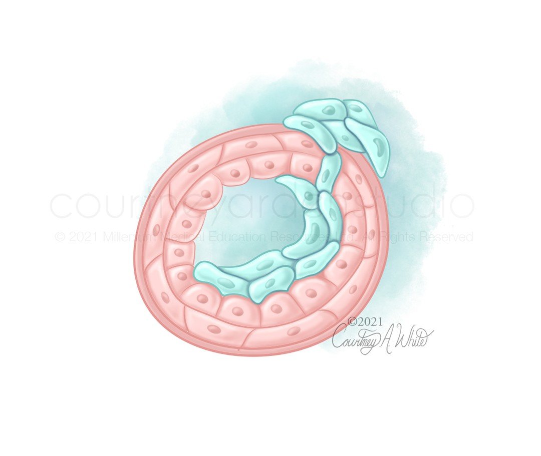
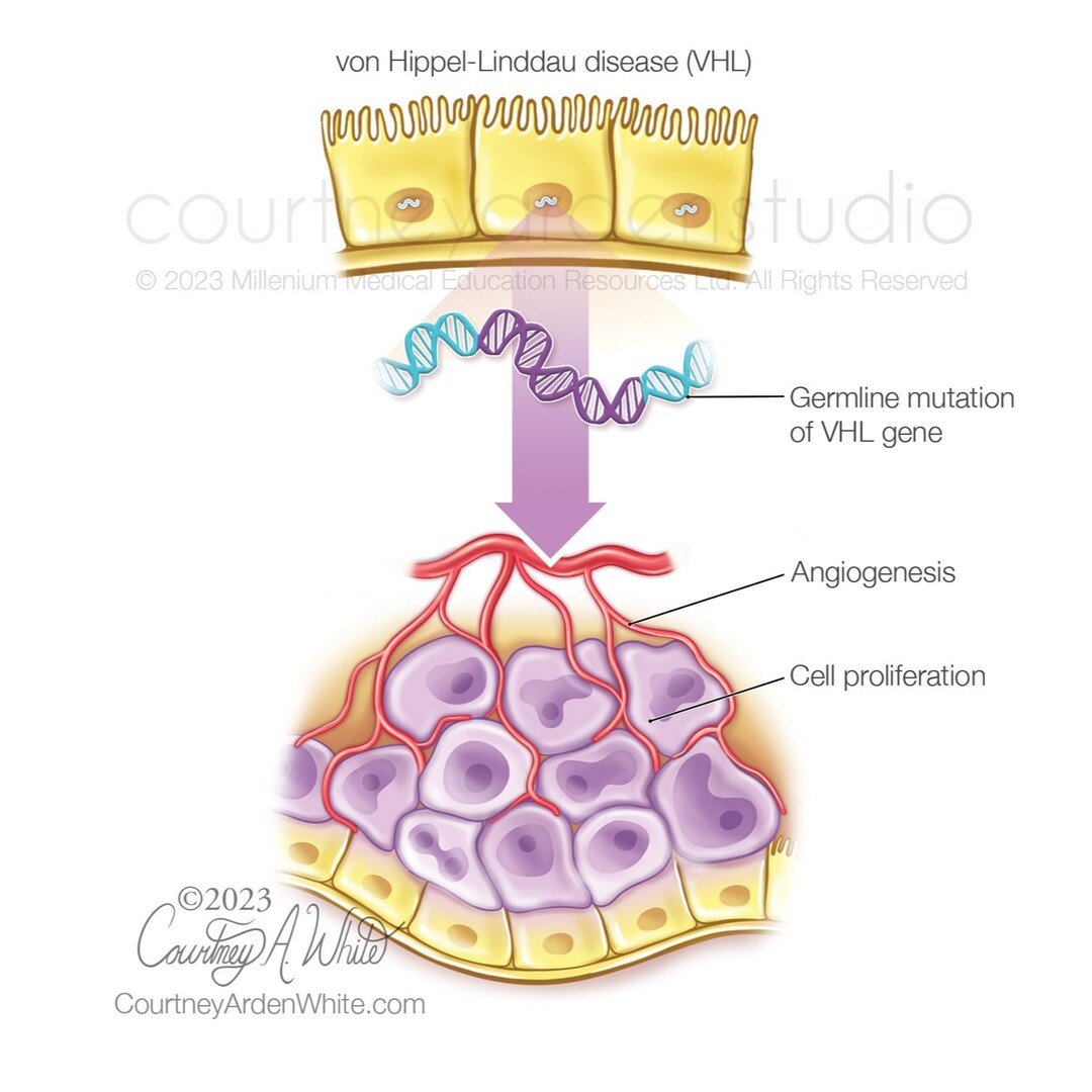
In a previous post, I shared a set of illustrations that were part of a patient education booklet about the kidneys and kidney diseases. These illustrations were a part of that same booklet. The first two illustrations explain von Hippel-Lindau (VHL) disease, which is a rare condition that causes tumors to grow in different parts of the body. This disease is caused by a mutation in the VHL gene and individuals with this gene mutation are at a higher risk of developing renal cell carcinoma (RCC). The third illustration shows how RCC starts in the cells that line the renal tubules. The fourth and final illustration depicts the Fuhrman grade scale, which is used to describe the aggressiveness of RCC cells and ranges from grade 1 to grade 4.
#medicalillustration #medart #medicalart #sciart #biologicalart #medicalcommunication #anatomyart #anatomicalart #artandanatomy #anatomydrawing #biologicalillustration #anatomyillustration #drawinganatomy #scienceart #scienceartist #scienceandart #medicalartist #medicaleducation #medicalstudent #meded #scicomm #scicommunity #healthcareart #patienteducation #vonhippellindaudisease #kidneydisease #kidneycancer #renalcellcarcinoma #cellularillustration #courtneyardenstudio